
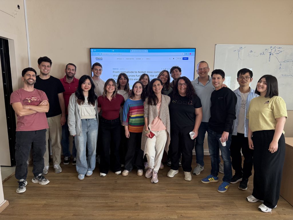

A recent study by scientists from the Medical School of the National and Kapodistrian University of Athens (NKUA) and the Institute of Biosciences and Applications (IBA) of the National Centre for Scientific Research Demokritos, in collaboration with researchers from the Biomedical Sciences Research Center Alexander Fleming, the University of Minho, and the University of Coimbra, has challenged the long-standing view that Tau is primarily important because of its role in Alzheimer’s disease pathology. The findings have been published in the premier international scientific journal Proceedings of the National Academy of Sciences (PNAS).
Tau is well-known for accumulating in the brains of patients with Alzheimer’s disease, where it forms pathological aggregates that contribute to neuronal dysfunction and cell death. However, Tau’s physiological role in healthy neurons has remained largely unknown. In the present study, researchers used both genetically modified mice and the nematode Caenorhabditis elegans to investigate Tau function and found that the absence of Tau leads to significant changes in mitochondrial morphology and function. Mitochondria are essential cellular organelles responsible for energy production and metabolic regulation.
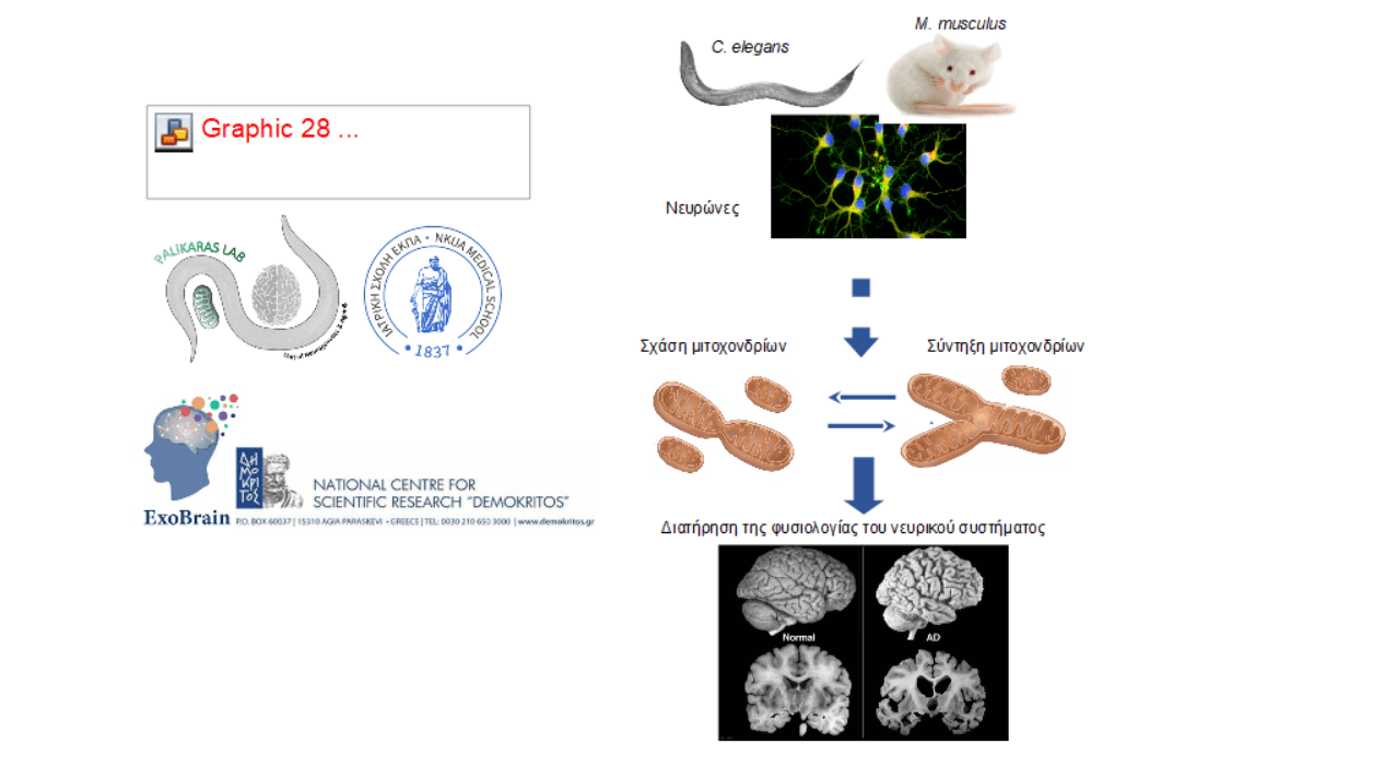
The research team, consisting of Dr. Eleni Tsakiri (Medical School, NKUA), Dr. Christina Ploumi (Medical School, NKUA), Kalliopi Skourti (IBA, Demokritos), Antonis Roussos (Medical School, NKUA), Eirini Mytilinaiou (Medical School, NKUA), and Dr. Anastasia Iakovou (IBA, Demokritos), headed by Dr. Ioannis Sotiropoulos (Researcher B, IBA, Demokritos) and Dr. Konstantinos Palikaras (Associate Professor, Medical School, NKUA), demonstrated that Tau deficiency promotes mitochondrial fusion, increases mitochondrial bioenergetic efficiency, and enhances quality-control mechanisms that protect neurons from damage.
As a result, neurons display increased resilience to metabolic and environmental stress. Importantly, the researchers also showed that these beneficial effects depend on mitofusins, a family of proteins that regulate mitochondrial fusion and morphology. Specifically, when mitofusin function was genetically abolished, the protective effects disappeared, demonstrating that mitochondrial morphology is a central mechanism through which Tau influences neuronal physiology.
For decades, the scientific community has focused primarily on what happens when Tau becomes mutated or pathological. This study reveals for the first time a fundamental physiological role of Tau in regulating mitochondrial function and neuronal adaptation to stress. These findings are particularly important because therapeutic strategies aimed at reducing Tau levels are currently being developed and tested in clinical trials for Alzheimer’s disease. A deeper understanding of Tau’s normal physiological functions is essential for designing safe and effective therapeutic interventions.
Overall, the study demonstrates for the first time that Tau is not merely a protein involved in neurodegeneration, but also a critical regulator of mitochondrial function, energy metabolism, and cellular survival. These results provide important new insights into the mechanisms that preserve neuronal health and may contribute to the development of novel therapeutic approaches for neurodegenerative disorders.
For additional information, please contact:
Associate Professor Konstantinos Palikaras
Head of the Research Unit of Neurogenetics and Ageing
Laboratory of Physiology
Medical School, National and Kapodistrian University of Athens (NKUA)
eMail: palikarask@med.uoa.gr
phone.: +302107462552

Dr. Ioannis Sotiropoulos, Researcher B
Director, ExoBrain Laboratory of Exosomal Analysis and Brain Pathology
Institute of Biosciences and Applications
National Centre for Scientific Research Demokritos
eMail: ioannis.@bio.demokritos.gr
phone.: +30210683588
Related links:


![Complete Anatomy Training Session [October 7, 2026]](https://hub.uoa.gr/en/wp-content/uploads/2026/10/NKUA_Invite-for-Complete-Anatomy-Training-Session_page-0001-1-1024x604.jpg)



![‘Dialogues for Athens’ Conference Organised by TO VIMA Under the Auspices of the National and Kapodistrian University of Athens and the Municipality of Athens [6-7 October]](https://hub.uoa.gr/en/wp-content/uploads/2026/10/Untitled-Design2-1-1024x576-1.jpg)